biofilm assay protocol
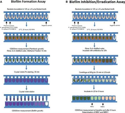
The protocol is divided into two assays that are performed independently to evaluate different activities of synthetic variants of HDPs. Workflow and timeline estimates for the biofilm inhibition gray boxes and eradication white boxes assays for testing the antibiofilm effects of.

Frontiers Biofilm And Planktonic Antibiotic Resistance In Patients With Acute Exacerbation Of Chronic Rhinosinusitis
Scanning Electron Microscopy SEM.

. Incubate the microtiter plate for 4-24 hrs at 37C. Its now understood to be the key reason why so many long-term health conditions persist and why antibiotics arent as effective as they once were. Cover assay plates and incubate at optimal growth temperature for desired amount of time.
Wipe the backside clean and fix biofilm formed at the air-liquid interface on the glass coverslip by immersing the coverslip in cold ethanol for 4 min or PFA for 30 min. Incubator 37C for 15 min then air-dry for 15 min. Aeruginosa biofilm was cultivated on 18 beads according to the standard protocol and processed as described above to determine the number of CFU per bead.
3 Set up four small trays in a series and add 1 to 2 inches of tap water to the last three. As part of the biofilm inhibition assay it. The results also demonstrate that cell viability generally correlates with biofilm biomass for-mation and that the CV2 method of biofilm measurement is probably more suitable than the CV1 method.
In addition some assays can. However the results in the tissue being. In summary the protocol optimized by ImQuest BioSciences provides a high throughput procedure to.
Center for Biofilm Engineering Paul Sturman PhD. Biofilm formation in microtiter plates is certainly the most commonly used method to grow and study biofilm. The biofilm processing was done as described above.
MBEC Assay Rotating Disk Reactor. Congo red agar CRA method that is a qualitative assay for detection of biofilm producer microorganism as a result of color change of colonies inoculated on CRA medium is described by Freeman et al. Stain all wells used in the assay with 125 μL of 01 crystal violet for 10 minutes.
This experiment was then repeated twice to achieve a total of three runs. Early phase biofilms are also prone to damage by the latter steps. Staining the Biofilm After incubation dump out cells by turning the plate over and shaking out the liquid.
Crystal violet CV assay is the most popular method for biofilm determination adopted by different laboratories so far. Trough base. Inoculate biofilm assay plates directly in 100-μl medium per well from the overnight microtiter plate cultures using a sterile 96-prong inoculating manifold.
In many assays biofilms are quantified by conventional culture plating method to get colony 10 forming unitscount which is an intensive procedure 1 Whereas other assays do use 96 well 11 microtiter plates for biofilm quantification as microtitre plate offers comparatively high 12. 1-2 Bacteria culture is prepared and dispensed into a 96-well microplate. In this method the negatively charged molecules within a biofilm.
This simple design is very popular due to its high-throughput screening capacities low cost and easy handling. 1 round-bottom 96-well plate 1 flat-bottom 96-well plate Appropriate media Appropriate antibiotic stock 15 mL conical tubes or glass test tubes for growing liquid cultures. Inoculate 5ml liquid medium with 5µl 1st Overnight culture use disposable test tubes and incubate at proper conditions overnight.
19162 100case. The quantitative assay methods for anti-biofilm activity based in colorimetric methodologies are similar to the microbroth dilution assay described in the Clinical Laboratory and Standards Institute CLSI document M7-A7. Wash 3 x 1 min in PBS Optional.
They use it to live and build colonies inside our bodies. Seal the plate with a cover or adhesive seal and incubate at a temperature of choice for 24-48 h. Rapid screening biofilm inhibition assay.
4 The peg lid is gently rinsed to removed planktonic bacteria and a serial diluted test solution is dispensed into a new 96-well microplate. Biofilm Assay Protocol for Biofilm assay by Safranin using 96-well plates. Its because these biofilms are nearly impossible to penetrate.
A step-by-step summary of the MBEC Assay. One of the first staining assays used in biofilm analysis was the crystal violet assay. Moderate shear - CSTR - ASTM Method E2196 Gentle shear - Batch - ASTM Method E2799.
General Biofilm Assay Protocol Materials Needed. Most protocols for embedding tissue describe placing the tissue flat in a histology tray placing a tissue cassette on top and covering in paraffin. For quantitative assays we typically use 4-8 replicate wells for each treatment.
Apply the second overnight culture into plates as your experimental designed pattern and perform the experiment as follow. In this assay the extent of biofilm formation is measured using the dye crystal violet CV. Products as well as biofilm testing protocols is not available to most industries as there has been minimal biocide testing for microorganisms in the biofilm state in the past.
- Shaking out the liquid wash one time with 200 ul dH2O and pipet out slowly to avoid disrupt the biofilm. In the protocol described here we focus on the use of 96-well optically clear polystyrene flat-bottom plate to study. MBEC Assay Biofilm Inoculator.
Shake the 96-well plate over. However biofilm layer formed at the liquid-air interphase known as pellicle is extremely sensitive to its washing and staining steps. Biofilm is a toxic slimy film that these bugs secrete.
Remove the glass coverslips from each well. 3 The peg lid is placed in the bacteria culture and incubated to generate the biofilm. In the protocol described here we will focus on the use of this assay to study biofilm formation by the model organism Pseudomonas aeruginosa.
The CRA medium is constructed by mixing 08 g of Congo red and 36 g of sucrose to 37 gL of Brain heart infusion BHI agar.

Biomolecules Free Full Text Critical Assessment Of Methods To Quantify Biofilm Growth And Evaluate Antibiofilm Activity Of Host Defence Peptides Html

Biofilm Formation Assay Kit Dojindo

An Overview Of The High Throughput Protocol For Metal Susceptibility Download Scientific Diagram

Polystyrene Microtiter Plate Biofilm Assay Of A Pleuropneumoniae Download Scientific Diagram

Microtiter Dish Biofilm Formation Assay Protocol

Biofilm Formation Assay Kit Dojindo Molecular Technologies Inc

Biofilm Viability Assay Kit Dojindo

Biofilm Formation Assay In Pseudomonas Syringae Bio Protocol

Schematic Crystal Violet Assay On Biofilms In A Microtiter Plate Download Scientific Diagram

Assess The Cell Viability Of Staphylococcus Aureus Biofilms Thermo Fisher Scientific Ng

Schematic Representation Of The Steps Involved In The Protocol For Download Scientific Diagram

Biofilm Formation Assay Kit Testpiece Dojindo Eu

Crystal Violet Assay To Assess The Antibiofilm Activity Of Samples Download Scientific Diagram

An Overview Of The High Throughput Protocol For Metal Susceptibility Download Scientific Diagram

A Combination Of Phenotype Microarraytm Technology With The Atp Assay Determines The Nutritional Dependence Of Escherichia Coli Biofilm Biomass Intechopen

Biofilm Eradication Testing For Antimicrobial Efficacy
0 Response to "biofilm assay protocol"
Post a Comment